Open reduction and internal fixation of metacarpal fractures using a thermoplastic splint as a surgical instrument
Article information
Abstract
Adequate positioning of the hand is a critical step in hand fracture operative repair that can impact both the clinical outcome and the efficiency of the operation. In this paper, we introduce the use of a thermoplastic splint with an added thumb stabilizing component as a means to increase the surgeon’s autonomy and to streamline the patient care pathway. The thermoplastic splint is custom fabricated preoperatively by the specialist hand therapist. The splint is used prior, during, and post operation with minimal modification. The thumb component assists maintaining the forearm in a stable pronated position whilst drilling and affixing metal work. This is demonstrated in the video of removal of metal work and open reduction and internal fixation of a metacarpal fracture.
INTRODUCTION
Fractures of the metacarpal (MC) bones account for a significant part of fractures in the hand; up to 40% as described in the literature with a shaft versus neck ratio of roughly 1:2 [1]. Multiple studies have shown that open reduction and internal fixation (ORIF) allows for early mobilization compared to intramedullary pinning fixation and is a frequent procedure performed for MC fractures [2,3].
Currently, the ORIF technique for MC fracture fixation is performed with the use of an assistant who holds the hand in a static pronated position for the operating surgeon, allowing ease of access to the fracture site. C-arm radiography is required during this procedure for visualization of fracture site and confirmation of metal work placement. Every time C-arm radiography is used the assistant must reposition the hand which is both time consuming and impairs the fluency of the procedure.
Some surgeons resort to a sterile crepe bandage or a “kidney” dish as a makeshift stand to help the assistant hold the hand in a fixed position. These solutions are frequently inadequate as the hand is repositioned multiple times throughout the procedure due to the natural tendency of the arm to supinate. Customized patient-precise instruments for hand surgery have been described before and are developed using additive manufacturing technology such as 3-dimensional printing, but there is no description of a device that inhibits the hand from supinating [4-6].
In our institution, all the patients with MC fractures that undergo ORIF will be followed up by our hand therapy colleagues and a thermoplastic splint will be custom fabricated based on the anatomy of the patient with variants upon the position of safe immobilization (POSI) [7-9].
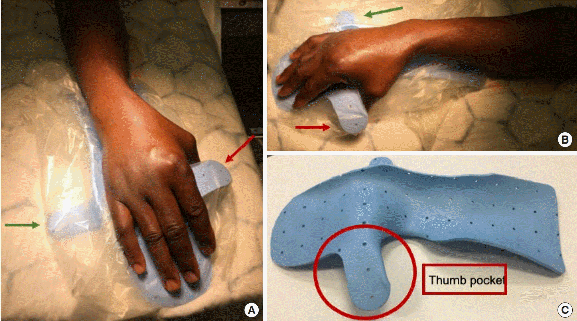
We developed a novel care pathway for patients deemed appropriate for MC ORIF fracture stabilization. The patients that fit the above characteristics when seen in the clinic are reviewed by the specialist hand therapist who measures and fabricates a modification of the POSI splint. This splint will be used preoperatively to immobilize the fracture as a customized stabilizing tool during the operation and then will serve as a resting splint postoperatively (Figs. 1, 2).

The modified thermoplastic splint. (A-C) The thumb pocket added to inhibit hand supination is shown with the red arrow. The additional layer of material which provides extra support is shown with the green arrow. The splint allows for the hand to lay naturally on the splint in pronation.
IDEA
The device was custom fabricated preoperatively molded directly on the patient to provide a suitable base of support for the hand. Final adjustments were made by the clinical specialist hand therapist at the time of fabrication to ensure appropriate positioning of the wrist and hand in a position of comfort; balancing tensions across joints and musculotendinous structures, in preference to James’ POSI [10]. This permitted the hand intrinsic muscles to be in a mid-range length minimizing load on the fracture site or through the metacarpophalangeal (MCP) joints. An integral component positioning the thumb in radial abduction was employed to assist maintaining forearm pronation when on the operating table (red arrow in Fig. 1). A removable piece of thermoplastic material is attached on the ulnar aspect of the splint providing further support to the hand and device limiting collapse when drilling the MC (green arrow in Fig. 1). Additionally, allowance was made in the splint for a potential increase in edema and the application of postoperative dressings.
Appropriate anatomic landmarks were obtained by the hand therapist using direct reference on the limb to determine dimensions of the device (Fig. 2). A 50% circumference of proximal forearm, distal anterior wrist crease and MCP joints were transferred to a template along with their respective longitudinal separation distances. Additional anatomic points were obtained; MCP obliquity to the wrist crease, the 2nd and 3rd digit webspace and the distance from the MCP to the tip of the 3rd digit [11]. These were again transferred to the template material, permitting development of a 2-dimensional representation of the device to be designed. The template was transferred to the thermoplastic stock and the material cut and subsequently softened in a heated hydrocollator. Material used: 3 mm (1/8”) perforated Rolyan Ezeform thermoplastic (Warrenville, IL, USA), heated to 75°C prior molding directly on the patient. The device was placed on the anterior aspect of the limb, permitting support for the digits, hand and wrist while allowing access for placement of surgical instrumentation and hardware. The material cured in several minutes to a rigid, radiolucent state. Due to the design of the splint that supports the forearm and prevents it from supination there is less tendency for the hand to supinate and therefore the material does not have to be heavy. In order to ascertain sterile field, the modified thermoplastic splint was inserted into a sterile transparent bag (Fig. 3).

Application of the device in surgery. (A) The thermoplastic splint used during the metacarpal open reduction and internal fixation procedure. (B) C-arm used while the stand is still in situ. (C) X-ray taken intraoperatively, providing evidence of radiolucency of the device (shadow of the splint seen on the upper left corner).
After the completion of surgery, the attached ulnar component was removed from the splint, patient placed in postoperative dressings and splint reapplied to rest the limb. Splint modification: removal of thumb component, was completed at the first postoperative therapy consultation. Hand therapy postoperative care was conducted in accord with the departmental clinical reasoning model described by Toemen and Midgley [12]. Based upon the UK market the average price for the material used for this splint is 70 GBP. No additional costs are incurred as customary postoperative hand therapy includes fabrication of a postoperative resting splint.
DISCUSSION
Achieving correct positioning of fracture site during surgery can play a critical role in surgical outcomes. This is particularly true of hand fractures where the combination of compact anatomy and the hand’s tendency to rotate supine at proximal joints can hinder optimal positioning of the fracture site for surgical intervention, including ORIF technique. Current practice often relies on a second surgeon or an assistant to hold the hand in the optimal position freeing up the lead surgeon to use both hands for operating. If an assistant is not available, or in addition to, the operating surgeon may use a makeshift stand using a sterile crepe bandage or a kidney dish to rest the hand on to keep the hand position in place.
The current practice as described requiring a second pair of hands for positioning means in resource scarce settings it is more difficult to achieve. Many countries have experienced staff shortages as clinicians were relocated to meet the changing demands of the 2020 COVID-19 (coronavirus disease 2019) pandemic. There is also an argument to be made in reducing the number of staff in the operating theatre; to reduce the number of people exposed to respiratory infections such as COVID-19 and having another clinician in the operating field can result in sharps injuries. The use of C-arm during the operation also exposes the assistant to radiation whilst stabilizing the hand.
The proposed innovation in this paper minimizes the aforementioned drawbacks whilst increasing the surgeon’s autonomy. This is achieved through stabilization of the hand at the thumb using a premolded personalized thermoplastic splint with an added thumb component. As shown in the Supplemental Video 1, the hand remains in pronation and stable during the procedure and can be achieved by a single experienced surgeon. The assisting surgeon can then perform traction while the hand is positioned on the splint to allow for fracture reduction. The increased autonomy not only improves the efficiency of the same operation but it also potentially frees up the second clinician to carry out a second operative list.
As well as the above-mentioned advantages during the operation, the thumb pocket splint also potentially reduces the number of times a patient is seen by the hand therapist in person, as the patient is able to carry on using the thermoplastic splint with the thumb pocket removed. This also eliminates the need for a temporary plaster of Paris splint pre-operation, a time and costsaving measure. By decreasing the need for postoperative appointments, we suspect a quicker discharge for the patient. Such simple operative measures can increase safety and efficiency and reduce cost of the treatment.
We have shown that with minor modifications, the traditional thermoplastic splint can be used as a stabilizing device during the MC fracture ORIF making the procedure more efficient and autonomous. The splint can then be modified and used postoperatively, making this process cost effective.
Notes
Conflict of interest
No potential conflict of interest relevant to this article was reported.
Ethical approval
The study was performed in accordance with the principles of the Declaration of Helsinki. Written informed consent was obtained.
Patient consent
The patients provided written informed consent for the publication and the use of their images.
Author contribution
Conceptualization: T Papavasiliou. Data curation: T Papavasiliou, PD Park, R Tejero, N Allain, L Uppal. Formal analysis: T Papavasiliou. Methodology: T Papavasiliou. Project administration: T Papavasiliou. Visualization: T Papavasiliou, N Allain. Writing - original draft: T Papavasiliou. Writing - review & editing: T Papavasiliou, PD Park, R Tejero, N Allain, L Uppal.
Supplementary Material
Supplemental Video 1. Use of the thermoplastic splint as a standing device on a case of periprosthetic metacarpal fracture open reduction and internal fixation. The material used for the splint demonstrated in this video is Woodcast based, the design is the same as shown in the figure. Supplemental data can be found at: https://doi.org/10.5999/aps.2021.00122.v001

